Mammogram
The Equipment
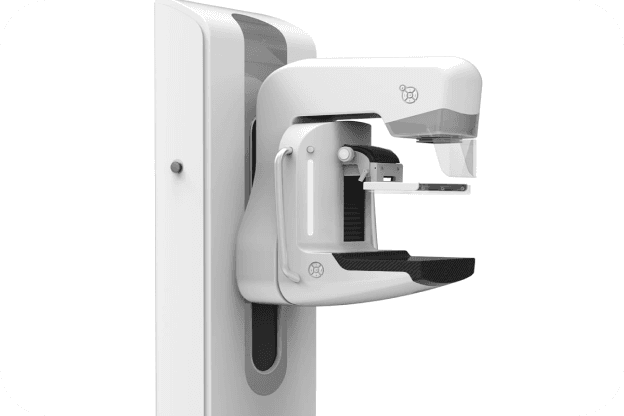
A mammography unit is a box with a tube that produces x-rays. The unit is used exclusively for breast x-ray exams and features special accessories to limit x-ray exposure to only the breast. The unit features a device to hold and compress the breast and position it so the technologist can capture images at different angles.
Breast tomosynthesis is performed using digital mammography units, but not all digital mammography machines are equipped to perform tomosynthesis imaging.
Summary
Mammography is specialized medical imaging that uses a low-dose x-ray system to see inside the breasts. A mammography exam, called a mammogram, aids in the early detection and diagnosis of breast diseases in women.
The exam is performed by placing your breast on a special platform and gradually compressed with a clear plastic paddle to take images. Preparation is needed day of the exam such as not wearing deodorant, talcum powder or lotion under your arms or on your breasts.
Please see below for details on the procedure, preparation, risks & benefits.
What is it
Three recent advances in mammography include digital mammography, computer-aided detection and breast tomosynthesis.
Digital mammography, also called full-field digital mammography (FFDM), is a mammography system in which the x-ray film is replaced by electronics that convert x-rays into mammographic pictures of the breast. These systems are similar to those found in digital cameras and their efficiency enables better pictures with a lower radiation dose. These images of the breast are transferred to a computer for review by the radiologist and for long term storage. The patient's experience during a digital mammogram is similar to having a conventional film mammogram.
Computer-aided detection (CAD) systems search digitized mammographic images for abnormal areas of density, mass, or calcification that may indicate the presence of cancer. The CAD system highlights these areas on the images, alerting the radiologist to carefully assess this area.
Breast tomosynthesis, also called three-dimensional (3-D) mammography and digital breast tomosynthesis (DBT), is an advanced form of breast imaging where multiple images of the breast from different angles are captured and reconstructed ("synthesized") into a three-dimensional image set. In this way, 3-D breast imaging is similar to computed tomography (CT) imaging in which a series of thin "slices" are assembled together to create a 3-D reconstruction of the body.
Although the radiation dose for some breast tomosynthesis systems is slightly higher than the dosage used in standard mammography, it remains within the FDA-approved safe levels for radiation from mammograms. Some systems have doses very similar to conventional mammography.
Large population studies have shown that screening with breast tomosynthesis results in improved breast cancer detection rates and fewer "call-backs," instances where women are called back from screening for additional testing because of a potentially abnormal finding.
Breast tomosynthesis may also result in:
Earlier detection of small breast cancers that may be hidden on a conventional mammogram
Fewer unnecessary biopsies or additional tests
Greater likelihood of detecting multiple breast tumors
Clearer images of abnormalities within dense breast tissue
Greater accuracy in pinpointing the size, shape and location of breast abnormalities
What is it used for?
Mammograms are used as a screening tool to detect early breast cancer in women experiencing no symptoms. They can also be used to detect and diagnose breast disease in women experiencing symptoms such as a lump, pain, skin dimpling or nipple discharge.
Screening Mammography
Mammography plays a central part in early detection of breast cancers because it can show changes in the breast years before a patient or physician can feel them. Current guidelines from the American College of Radiology (ACR) and the National Comprehensive Cancer Network (NCCN) recommend screening mammography every year for women, beginning at age 40. Research has shown that annual mammograms lead to early detection of breast cancers, when they are most curable and breast-conservation therapies are available.
The ACR and the National Cancer Institute (NCI) also suggest that women who have had breast cancer, and those who are at increased risk due to a family history of breast or ovarian cancer, should seek expert medical advice about whether they should begin screening before age 40 and the need for other types of screening. If you are at high risk for breast cancer, you may need to obtain a breast MRI in addition to your annual mammogram.
Diagnostic Mammography
Diagnostic mammography is used to evaluate a patient with abnormal clinical findings—such as a breast lump or nipple discharge—that have been found by the woman or her doctor. Diagnostic mammography may also be done after an abnormal screening mammogram in order to evaluate the area of concern on the screening exam.
How to Prepare
Before scheduling a mammogram, the American Cancer Society (ACS) and other specialty organizations recommend that you discuss any new findings or problems in your breasts with your doctor. In addition, inform your doctor of any prior surgeries, hormone use, and family or personal history of breast cancer.
Do not schedule your mammogram for the week before your menstrual period if your breasts are usually tender during this time. The best time for a mammogram is one week following your period. Always inform your doctor or x-ray technologist if there is any possibility that you are pregnant.
The ACS also recommends you:
Do not wear deodorant, talcum powder or lotion under your arms or on your breasts on the day of the exam. These can appear on the mammogram as calcium spots.
Describe any breast symptoms or problems to the technologist performing the exam.
Obtain your prior mammograms and make them available to the radiologist if they were done at a different location. This is needed for comparison with your current exam and can often be obtained on a CD.
Ask when your results will be available; do not assume the results are normal if you do not hear from your doctor or the mammography facility.
How the test is performed
Mammography is performed on an outpatient basis.
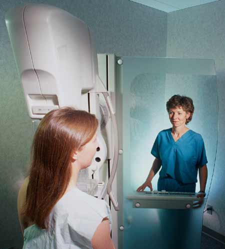
During mammography, a specially qualified radiologic technologist will position your breast in the mammography unit. Your breast will be placed on a special platform and compressed with a clear plastic paddle. The technologist will gradually compress your breast.
Breast compression is necessary in order to:
Even out the breast thickness so that all of the tissue can be visualized.
Spread out the tissue so that small abnormalities are less likely to be hidden by overlying breast tissue.
Allow the use of a lower x-ray dose since a thinner amount of breast tissue is being imaged.
Hold the breast still in order to minimize blurring of the image caused by motion.
Reduce x-ray scatter to increase sharpness of picture.
You will be asked to change positions between images. The routine views are a top-to-bottom view and an angled side view. The process will be repeated for the other breast. Compression is still necessary for tomosynthesis imaging in order to minimize motion, which degrades the images. During screening breast tomosynthesis, two-dimensional images are also obtained or created from the synthesized 3-D images.
You must hold very still and may need to hold your breath for a few seconds while the technologist takes the x-ray. This helps reduce the possibility of a blurred image. The technologist will walk behind a wall or into the next room to activate the x-ray machine.
When the examination is complete, the technologist may ask you to wait until the radiologist confirms they have all the necessary images.
The examination process should take about 30 minutes.
What it feels like
You will feel pressure on your breast as it is squeezed by the compression paddle. Some women with sensitive breasts may experience discomfort. If this is the case, schedule the procedure when your breasts are least tender. Be sure to inform the technologist if pain occurs as compression is increased. If discomfort is significant, less compression will be used. Always remember compression allows better quality mammograms.
Getting results
A radiologist, a doctor trained to supervise and interpret radiology examinations, will analyze the images. The radiologist will send a signed report to your primary care or referring physician who will discuss the results with you.
You will also be notified of the results by the mammography facility.
Call Backs
You may need a follow-up exam. If so, your doctor will explain why. Sometimes a follow-up exam further evaluates a potential issue with more views or a special imaging technique. It may also see if there has been any change in an issue over time. Follow-up exams are often the best way to see if treatment is working or if a problem needs attention.
Benefits
Screening mammography reduces the risk of death due to breast cancer. It is useful for detecting all types of breast cancer, including invasive ductal and invasive lobular cancer.
Screening mammography improves a physician's ability to detect small tumors. When cancers are small, the woman has more treatment options.
The use of screening mammography increases the detection of small abnormal tissue growths confined to the milk ducts in the breast, called ductal carcinoma in situ (DCIS).
No radiation stays in your body after an x-ray exam.
X-rays usually have no side effects in the typical diagnostic range for this exam.
Risks
There is always a slight chance of cancer from excessive exposure to radiation. However, given the small amount of radiation used in medical imaging, the benefit of an accurate diagnosis far outweighs the associated risk.
The radiation dose for this procedure varies. See the Radiation Dose page for more information.
False Positive Mammograms. Five percent to 15 percent of screening mammograms require more testing such as additional mammograms or ultrasound. Most of these tests turn out to be normal. If there is an abnormal finding, a follow-up or biopsy may have to be performed. Most of the biopsies confirm that no cancer was present. It is estimated that a woman who has yearly mammograms between ages 40 and 49 has about a 30 percent chance of having a false-positive mammogram at some point in that decade and about a 7 percent to 8 percent chance of having a breast biopsy within the 10-year period.
Women should always tell their doctor and x-ray technologist if they are pregnant. See the Radiation Safety page for more information about pregnancy and x-rays.
The Equipment
A mammography unit is a box with a tube that produces x-rays. The unit is used exclusively for breast x-ray exams and features special accessories to limit x-ray exposure to only the breast. The unit features a device to hold and compress the breast and position it so the technologist can capture images at different angles.
Breast tomosynthesis is performed using digital mammography units, but not all digital mammography machines are equipped to perform tomosynthesis imaging.
Let Us Help You Prepare For Your Scan
We do the leg work of assembling scan information from your test facility and other reputable sources to give women a complete picture.
For More Information About Mammogram
Phone: 800-227.-2345
National Breast Cancer Foundation, Inc.
Phone: 972-248-9200
Susan G. Komen Breast Cancer Foundation
Phone: 1-877 GO KOMEN