Breast Ultrasounds
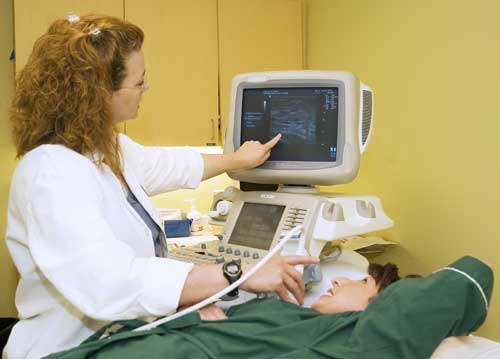
The Equipment
Ultrasound machines consist of a computer console, video monitor and an attached transducer. The transducer is a small hand-held device that resembles a microphone. Some exams may use different transducers (with different capabilities) during a single exam. The transducer sends out inaudible, high-frequency sound waves into the body and listens for the returning echoes. The same principles apply to sonar used by boats and submarines.
The technologist applies a small amount of gel to the area under examination and places the transducer there. The gel allows sound waves to travel back and forth between the transducer and the area under examination. The ultrasound image is immediately visible on a video monitor. The computer creates the image based on the loudness (amplitude), pitch (frequency), and time it takes for the ultrasound signal to return to the transducer. It also considers what type of body structure and/or tissue the sound is traveling through.
Summary
Ultrasound imaging of the breast uses sound waves to produce pictures of the internal structures of the breast. It is used to help diagnose breast lumps or other abnormalities found during a physical exam, or on a mammogram or breast MRI.
Ultrasound is safe, noninvasive, and does not use radiation. You will lie on the exam table and maybe asked to raise your arm(s) above your head. The sonographer will apply a water-based gel to the area under examination and place the transducer on the body and move it back and forth over the area of interest until it captures the desired images.
This exam requires little to no special preparation. Leave jewelry at home and wear loose, comfortable clothing. You will need to undress from the waist up and to wear a gown during the exam.
Please see below for details on the procedure, preparation, risks & benefits.
What is it
Ultrasound imaging is a noninvasive medical test that helps physicians diagnose and treat medical conditions. It is safe and painless. It produces pictures of the inside of the body using sound waves. Ultrasound imaging is also called sonography. It uses a small probe called a transducer and gel placed directly on the skin. High-frequency sound waves travel from the probe through the gel into the body. The probe collects the sounds that bounce back. A computer uses those sound waves to create an image. Ultrasound exams do not use radiation (x-rays). Because ultrasound captures images in real-time, it can show the structure and movement of the body's internal organs. The images can also show blood flowing through blood vessels.
Doppler ultrasound is a special ultrasound technique that evaluates movement of materials in the body. It allows the doctor to see and evaluate blood flow through arteries and veins in the body.
Ultrasound (US) of the breast produces a picture of the internal structures of the breast.
During a breast ultrasound exam, the sonographer or doctor may use Doppler techniques to evaluate blood flow or lack of flow in any breast mass. In some cases, this may provide additional information as to the cause of the mass.
How it works
Ultrasound imaging uses the same principles as the sonar that bats, ships, and fishermen use. When a sound wave strikes an object, it bounces back or echoes. By measuring these echo waves, it is possible to determine how far away the object is as well as its size, shape, and consistency. This includes whether the object is solid or filled with fluid.
Doctors use ultrasound to detect changes in the appearance of organs, tissues, and vessels and to detect abnormal masses, such as tumors.
In an ultrasound exam, a transducer both sends the sound waves and records the echoing (returning) waves. When the transducer is pressed against the skin, it sends small pulses of inaudible, high-frequency sound waves into the body. As the sound waves bounce off internal organs, fluids and tissues, the sensitive receiver in the transducer records tiny changes in the sound's pitch and direction. A computer instantly measures these signature waves and displays them as real-time pictures on a monitor. The technologist typically captures one or more frames of the moving pictures as still images. They may also save short video loops of the images.
Doppler ultrasound, a special ultrasound technique, measures the direction and speed of blood cells as they move through vessels. The movement of blood cells causes a change in pitch of the reflected sound waves (called the Doppler effect). A computer collects and processes the sounds and creates graphs or color pictures that represent the flow of blood through the blood vessels.
How to prepare
You will need to undress from the waist up and to wear a gown during the exam.
How the test is performed
You will lie on your back or on your side on the exam table. The sonographer may ask you to raise your arm(s) above your head.
The radiologist (a doctor specifically trained to supervise and interpret radiology exams) or sonographer will position you on the exam table. They will apply a water-based gel to the area of the body under examination. The gel will help the transducer make secure contact with the body. It also eliminates air pockets between the transducer and the skin that can block the sound waves from passing into your body. The sonographer places the transducer on the body and moves it back and forth over the area of interest until it captures the desired images.
There is usually no discomfort from pressure as they press the transducer against the area being examined. However, if the area is tender, you may feel pressure or minor pain from the transducer.
Doctors perform Doppler sonography with the same transducer.
Once the imaging is complete, the technologist will wipe off the clear ultrasound gel from your skin. Any portions that remain will dry quickly. The ultrasound gel does not usually stain or discolor clothing.
What it feels like
Most ultrasound exams are painless, fast, and easily tolerated.
Breast ultrasound is usually completed within 30 minutes.
If the doctor performs a Doppler ultrasound exam, you may hear pulse-like sounds that change in pitch as they monitor and measure the blood flow.
You may need to change positions during the exam.
When the exam is complete, the technologist may ask you to dress and wait while they review the ultrasound images.
After an ultrasound exam, you should be able to resume your normal activities immediately.
Getting results
A radiologist, a doctor trained to supervise and interpret radiology exams, will analyze the images. The radiologist will send a signed report to the doctor who requested the exam. Your doctor will then share the results with you. In some cases, the radiologist may discuss results with you after the exam.
You may need a follow-up exam. If so, your doctor will explain why. Sometimes a follow-up exam further evaluates a potential issue with more views or a special imaging technique. It may also see if there has been any change in an issue over time. Follow-up exams are often the best way to see if treatment is working or if a problem needs attention.
Benefits
Most ultrasound scanning is noninvasive (no needles or injections).
Occasionally, an ultrasound exam may be temporarily uncomfortable, but it should not be painful.
Ultrasound is widely available, easy to use, and less expensive than most other imaging methods.
Ultrasound imaging is extremely safe and does not use radiation.
Ultrasound scanning gives a clear picture of soft tissues that do not show up well on x-ray images.
Ultrasound provides real-time imaging. This makes it a good tool for guiding minimally invasive procedures such as needle biopsies and fluid aspiration.
Ultrasound imaging can help detect lesions in women with dense breasts.
Ultrasound may help detect and classify a breast lesion that cannot be interpreted adequately through mammography alone.
Using ultrasound, doctors are able to determine that many areas of clinical concern are due to normal tissue (such as fat lobules) or benign cysts. For most women 30 years of age and older, a mammogram will be used together with ultrasound. For women under age 30, ultrasound alone is often enough to determine whether an area of concern needs a biopsy or not.
Risks
Standard diagnostic ultrasound has no known harmful effects on humans.
Interpretation of a breast ultrasound exam may lead to additional procedures such as follow-up ultrasound and/or aspiration or biopsy. Many of the areas thought to be of concern turn out to be non-cancerous (false positives).
The Equipment
Ultrasound machines consist of a computer console, video monitor and an attached transducer. The transducer is a small hand-held device that resembles a microphone. Some exams may use different transducers (with different capabilities) during a single exam. The transducer sends out inaudible, high-frequency sound waves into the body and listens for the returning echoes. The same principles apply to sonar used by boats and submarines.
The technologist applies a small amount of gel to the area under examination and places the transducer there. The gel allows sound waves to travel back and forth between the transducer and the area under examination. The ultrasound image is immediately visible on a video monitor. The computer creates the image based on the loudness (amplitude), pitch (frequency), and time it takes for the ultrasound signal to return to the transducer. It also considers what type of body structure and/or tissue the sound is traveling through.
Let Us Help You Prepare For Your Scan
We do the leg work of assembling scan information from your test facility and other reputable sources to give women a complete picture.
For More Information About Breast Ultrasounds
Phone: 800-227.-2345
National Breast Cancer Foundation, Inc.
Phone: 972-248-9200
Susan G. Komen Breast Cancer Foundation
Phone: 1-877 GO KOMEN